Collagen remodeling and increased type I collagen density following non-ablative monopolar radiofrequency for vulvar laxity: A prospective pilot study
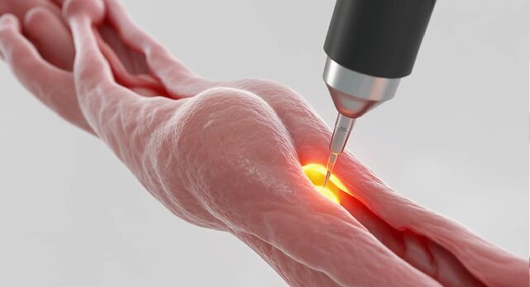
The aim of this study was to evaluate the clinical and histological effects of a single session of non-ablative monopolar radiofrequency on vulvar laxity, with emphasis on collagen remodeling.
Highlights
- •Non-ablative monopolar radio-frequency improved vulvar tissue laxity.
- •Histology showed increased type I collagen after treatment.
- •Patients reported higher satisfaction and improved vulvar firmness.
Abstract
Introduction
Vulvar laxity is associated with alterations in dermal extracellular matrix structure, particularly collagen organization, resulting in reduced tissue firmness and elasticity. Energy-based technologies have been proposed as minimally invasive approaches for tissue remodeling. The aim of this study was to evaluate the clinical and histological effects of a single session of non-ablative monopolar radiofrequency on vulvar laxity, with emphasis on collagen remodeling.
Methods
This prospective study included nine women aged 30–60 years with vulvar laxity. Vulvar biopsies were obtained before treatment (T0) and at 90 days (T3). Participants underwent a single session of non-ablative monopolar radiofrequency (6 MHz, 60 % power), with dermal temperature maintained at 45–60 °C and epidermal cooling. Follow-up was performed immediately and at 30, 90 and 180 days. Collagen types I and III were quantified using Picro-Sirius Red under polarized light. Data were analyzed using the paired Wilcoxon test (p < 0.05).
Results
Significant improvements were observed in all patient-reported outcomes at 90 days. Mean scores increased from 2.3 to 4.2 for perceived vulvar laxity, 2.7 to 4.3 for overall vulvar appearance, 2.1 to 4.2 for skin firmness, and 2.3 to 4.3 for comfort with genital appearance (all p < 0.05). Pinch Test measurements decreased significantly from baseline, indicating reduced skin fold thickness, with effects maintained throughout the 180-day follow-up (p < 0.05). Histologically, type I collagen increased from 8,986.65 ± 8,006.84 to 19,545.96 ± 4,570.01 µm2 (p = 0.038), type III collagen decreased from 15,779.00 ± 3,980.40 to 8,626.27 ± 1,263.47 µm2 (p = 0.017), and the collagen I/III ratio increased from 0.33 ± 0.50 to 1.77 ± 0.44 (p = 0.011), indicating collagen remodeling.
Conclusion
Non-ablative monopolar radiofrequency promoted clinical improvement and collagen remodeling in vulvar tissue, supporting its role as a minimally invasive option for vulvar laxity.
Introduction
Vulvar laxity results from alterations in the extracellular matrix (ECM), influenced by factors such as hormonal changes, skin aging, pregnancies, weight fluctuations, and mechanical stress that compromise the architecture of vulvar skin [1], [2], [3].
Within the ECM, type I collagen is the main structural component of the dermis, associated with tissue strength and support, whereas type III collagen contributes to elasticity and tissue remodeling. Elastic fibers provide tissue distensibility, and changes in the quantity and organization of these structures may lead to reduced skin firmness and elasticity, contributing to vulvar laxity [2], [3].
In recent years, there has been an increased demand for treatments focused on vulvar health, reflecting greater attention to female genital comfort and aesthetic concerns [4]. In this context, energy-based technologies have been used to promote tissue remodeling [5], [6]. Non-ablative monopolar radiofrequency induces controlled heating in the deep dermal layers, stimulating neocollagenesis, neoelastogenesis, and ECM reorganization [7], [8].
Recent studies have demonstrated benefits of radiofrequency in the treatment of female genital laxity, including improvements in tissue firmness and patient satisfaction [9]. However, objective histological evidence regarding the impact of monopolar radiofrequency on vulvar collagen organization remains limited.
Histological analysis allows the evaluation of structural modifications following therapeutic interventions and contributes to understanding the mechanisms involved in tissue response [10].
This study aimed to evaluate the clinical, histological, and subjective efficacy of non-ablative monopolar radiofrequency with automatic cooling in the treatment of vulvar laxity in adult women, by comparing parameters before and after the intervention.
Methods
The study sample initially consisted of ten women aged between 30 and 60 years with vulvar laxity who met the study eligibility criteria. However, one participant was excluded after becoming pregnant during the follow-up period, resulting in a final sample of nine women. Sample collection was performed at NGR Clínica Médica LTDA, while histological processing was conducted at the Federal University of Paraíba, in João Pessoa, PB, Brazil.
This study was approved by the institutional Research Ethics Committee (n° 91697625.0.0000.5188).
Participants were evaluated at five time points: baseline (T0), immediate (T1), 30 days (T2), 90 days (T3), and 180 days (T4). At baseline (T0), a detailed anamnesis and complete gynecological physical examination were performed.
For clinical assessment of vulvar laxity, the methodology proposed by Guedes-Batalha, as described by Carvalho et al. [11], was used, including the pinch test to measure the degree of tissue laxity. This classification categorizes vulvar laxity into four grades based on severity and the target structures involved (skin, fat, or both), facilitating standardized clinical evaluation and treatment planning.
Standardized photographs of the vulvar region were taken to allow intra-individual comparison throughout follow-up. Participants completed a subjective genital self-image questionnaire using a Likert scale from 0 to 5, where higher values indicate greater satisfaction with intimate appearance. Additionally, a baseline vulvar biopsy was performed under local anesthesia, with tissue samples collected for histopathological analysis prior to treatment.
At baseline (T0), a structured data collection proforma developed by the authors was also applied to characterize sociodemographic and clinical variables, including continuous variables (age, body weight, and number of pregnancies) and categorical variables (ethnicity, menopausal status, physical activity level, weight fluctuation ≥ 5 kg per year, urinary incontinence, vaginal dryness, and discomfort during sexual intercourse). Physical activity level was categorized according to weekly frequency.
The second stage consisted of a single therapeutic intervention using non-ablative monopolar radiofrequency, with the Coolfase® device (IBRAMED, Amparo, SP, Brazil), operating at 6 MHz and 60 % power, using a 4 cm2 metal applicator in continuous mode. Dermal temperature was maintained between 45 and 60 °C with continuous monitoring, while automatic cooling preserved the epidermis between 40 and 42 °C. The protocol included 10 passes per point in the treated area, with a mean of 215.4 shots per patient.
The third stage consisted of longitudinal follow-up, with clinical and photographic reassessments performed immediately after the procedure (T1), and at 30 (T2), 90 (T3), and 180 days (T4). The subjective genital self-image questionnaire was applied before the procedure (T0) and repeated at 90 days (T3), using the same scale as at baseline. At this same time point (T3), a new biopsy was performed for histopathological analysis. Biopsy sites were standardized and collected from the medial aspect of the labium majus, adjacent to the interlabial sulcus, at a point equidistant between the clitoral hood and the vaginal introitus. To avoid sampling scar tissue from the initial procedure, the second biopsy was obtained from the contralateral hemivulva at the corresponding anatomical location.
Histopathological and histomorphometric evaluation
Vulvar tissue samples (T0 and T3) were fixed in 10 % neutral buffered formalin for 48 h at room temperature (23 ± 2 °C). Subsequently, samples were processed using standard histological procedures, including dehydration in graded ethanol (70 %, 80 %, 90 %, and 100 %), clearing in xylene, and paraffin embedding (maximum temperature of 60 °C). Serial sections (4 µm thick) were obtained, mounted on glass slides, deparaffinized, and rehydrated through decreasing ethanol concentrations.
Sections were stained with hematoxylin and eosin (H&E) and Picro Sirius Red. Histological evaluation was performed using a Nikon Eclipse Ci-L light microscope (Nikon, Tokyo, Japan) at 40 × magnification, coupled with a Nikon DS-Ri2 digital camera. Image acquisition and processing were carried out using NIS-Elements D software (version 4.00, Nikon).
For collagen analysis, Picro Sirius Red-stained sections were examined under polarized light, allowing differentiation of collagen fibers based on birefringence properties, with type I collagen appearing in red–orange hues and type III collagen in greenish tones.
Quantitative analysis was performed by converting images into binary format, followed by measurement of collagen fiber areas (µm2). Color segmentation was applied to identify red, yellow, and green pixels corresponding to collagen fibers. Measurements were performed using Image-Pro Plus software (Media Cybernetics, Rockville, MD, USA).
Statistical analysis
Histomorphometric data (type I collagen, type III collagen, total collagen, and I/III ratio) were compared between baseline (T0) and post-treatment (T3) using the paired Wilcoxon test.
The pinch test (vulvar skin fold thickness, in centimeters) was evaluated at T0, T1, T2, T3, and T4, with data expressed as median and range. For repeated measures analysis, the Friedman test was used, followed by the Wilcoxon test for pairwise comparisons between time points. A significance level of 5 % (p < 0.05) was adopted. Statistical analyses were performed using IBM SPSS Statistics (IBM Corp., Armonk, NY, USA), and tables were created using Microsoft Excel.
Results
Clinical outcomes
The mean age of participants was 44.0 ± 11.6 years, with a mean body weight of 64.4 ± 8.5 kg. Regarding clinical characteristics, 33.3 % were menopausal and 66.7 % premenopausal. Regular physical activity was reported by 55.6 % of participants. The most prevalent gynecological complaints were urinary incontinence and vaginal dryness, both observed in 77.8 % of the cohort (Table 1).
Table 1. Sociodemographic, clinical characteristics and gynecological complaints of participants.
| Characteristic | |
|---|---|
| Mean ± SD | |
| Age (years) | 44.0 ± 11.6 |
| Weight (kg) | 64.4 ± 8.5 |
| Number of pregnancies | 2.4 ± 1.6 |
| N (%) | |
| Ethnicity | |
| White | 5 (55.6) |
| Mixed race | 4 (44.4) |
| Menopause | |
| Yes | 3 (33.3) |
| No | 6 (66.7) |
| Physical activity | |
| None | 2 (22.2) |
| 1–2x/week | 2 (22.2) |
| ≥3x/week | 5 (55.6) |
| Weight fluctuation | |
| Yes | 1 (12.5) |
| No | 8 (87.5) |
| Urinary incontinence | |
| Yes | 7 (77.8) |
| No | 2 (22.2) |
| Vaginal dryness | |
| Yes | 7 (77.8) |
| No | 2 (22.2) |
| Dyspareunia/discomfort | |
| Yes | 4 (44.4) |
| No | 5 (55.6) |
Data are expressed as mean ± standard deviation or number (percentage). SD: standard deviation.
In the evaluation of participant satisfaction and clinical outcomes, an increase in mean scores was observed after treatment (T3) compared to baseline (T0) for all assessed parameters (Table 2). The perceived level of vulvar laxity improved from 2.3 at baseline to 4.2 after treatment; overall appearance of the intimate skin region improved from 2.7 to 4.3; vulvar skin firmness improved from 2.1 to 4.2; and comfort and well-being regarding intimate appearance improved from 2.3 to 4.3. The Wilcoxon test demonstrated statistically significant differences for all outcomes (p < 0.05).
Table 2. Patient satisfaction assessment and clinical outcomes before and after treatment (N = 9).
| Parameter | T0 (mean ± SD) | T3 (mean ± SD) | p-value |
|---|---|---|---|
| Perceived laxity | 2.3 ± 1.4 | 4.2 ± 0.8 | 0.01 |
| Overall appearance of intimate área | 2.7 ± 1.1 | 4.3 ± 0.7 | 0.04 |
| Vulvar skin firmness | 2.1 ± 1.3 | 4.2 ± 0.7 | 0.01 |
| Comfort and well-being | 2.3 ± 1.5 | 4.3 ± 0.5 | 0.01 |
Scores were assessed using a 0–5 scale, with higher values indicating greater intensity.
Data are expressed as mean ± SD.
Wilcoxon signed-rank test was used for paired comparisons between T0 and T3.
p < 0.05 was considered statistically significant.
Pinch test results
The Friedman test demonstrated a significant difference among the evaluated time points (χ2 = 17.583; p = 0.001), indicating variation in clinical scores over time. A significant reduction in Pinch Test measurements was observed after baseline (T0), indicating a decrease in skin fold thickness and clinical improvement following the intervention.
The Pinch Test measurements showed a progressive reduction over time, with median values of 4.0 (3.0–6.0) cm at T0, 3.5 (2.0–5.0) cm at T1, 3.0 (3.0–5.0) cm at T2, 3.5 (2.0–4.5) cm at T3, and 4.0 (2.0–5.0) cm at T4 (Table 3). In pairwise comparisons using the Wilcoxon test, significant differences were observed between T0 and T1 (p = 0.008), T0 and T2 (p = 0.014), T0 and T3 (p = 0.007), and T0 and T4 (p = 0.026). (Table 3). No significant differences were found among post-intervention time points, suggesting maintenance of the effect throughout the follow-up period, in agreement with the visual findings presented in Fig. 1.
Table 3. Evolution of vulvar Pinch Test measurements during follow-up (N = 9).
| Time point | Median (range) (cm) | p-value |
|---|---|---|
| T0 | 4.0 (3.0 – 6.0) | – |
| T1 | 3.5 (2.0 – 5.0) | 0.008 |
| T2 | 3.0 (3.0 – 5.0) | 0.014 |
| T3 | 3.5 (2.0 – 4.5) | 0.007 |
| T4 | 4.0 (2.0 – 5.0) | 0.026 |
Pinch test: measurement of vulvar skin fold thickness, expressed in centimeters.
T0: baseline; T1: immediate post-procedure; T2: 30 days; T3: 90 days; T4: 180 days after treatment.
Data are expressed as median (range).
Friedman test was used for repeated measures, followed by Wilcoxon signed-rank test for pairwise comparisons.
p-values refer to comparisons with baseline (T0).p < 0.05 was considered statistically significant.

Histological findings
Histological evaluation revealed dermal stromal remodeling between baseline (T0) and 90 days after the procedure (T3), characterized by collagen fiber reorganization and increased structural density (Fig. 2). At T0, qualitative analysis revealed a weakened collagen pattern, with short, fragmented, and disorganized bundles. The reticular dermis showed low fiber density and clear interstitial spaces, suggesting a loose collagen network. Quantitatively, this period was characterized by a predominance of type III collagen (15,779.00 ± 3,980.40 µm2) compared to type I collagen (8,986.65 ± 8,006.84 µm2), resulting in a low Col I/III ratio (0.33 ± 0.50) (Table 4).
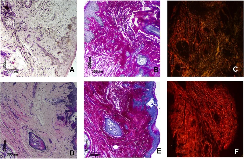
Fig. 2. Histological images obtained at baseline (T0: A–C) and after treatment (T3: D–F). A and D: hematoxylin and eosin (H&E); B and E: Picrosirius Red under bright-field microscopy; C and F: Picrosirius Red under polarized light. At T0, the tissue exhibited predominance of type III collagen, with lower type I collagen density and a loosely organized fibrillar pattern. At T3, increased type I collagen and reduced type III collagen were observed, with a denser and more organized collagen architecture, indicating extracellular matrix maturation. Magnification: ×40; scale bar: 300 µm. (For interpretation of the references to colour in this figure legend, the reader is referred to the web version of this article.)
Table 4. Collagen content before and after non-ablative monopolar radiofrequency treatment.
| Variable | Before (mean ± SD) | After (mean ± SD) | p-value |
|---|---|---|---|
| Collagen I (µm2) | 8,986.65 ± 8,006.84 | 19,545.96 ± 4,570.01 | 0.038 |
| Collagen III (µm2) | 15,779.00 ± 3,980.40 | 8,626.27 ± 1,263.47 | 0.017 |
| Collagen I/III ratio | 0.33 ± 0.50 | 1.77 ± 0.44 | 0.011 |
| Total collagen | 12,382.83 ± 7,059.56 | 13,638.55 ± 6,400.23 | 0.744 |
Data are expressed as mean ± standard deviation.Comparisons between before and after treatment were performed using the Wilcoxon signed-rank test.
p < 0.05 was considered statistically significant.
After 90 days (T3), morphological changes in tissue architecture were observed. There was a significant increase in type I collagen deposition from 8,986.65 ± 8,006.84 µm2 at T0 to 19,545.96 ± 4,570.01 µm2 at T3 (p = 0.038). In parallel, type III collagen showed a statistically significant reduction from 15,779.00 ± 3,980.40 µm2 at T0 to 8,626.27 ± 1,263.47 µm2 at T3 (p = 0.017). This variation was reflected in an increase in the Col I/III ratio from 0.33 ± 0.50 at T0 to 1.77 ± 0.440 at T3 (p = 0.011), suggesting an ECM with characteristics compatible with a higher degree of structural organization (Fig. 3).
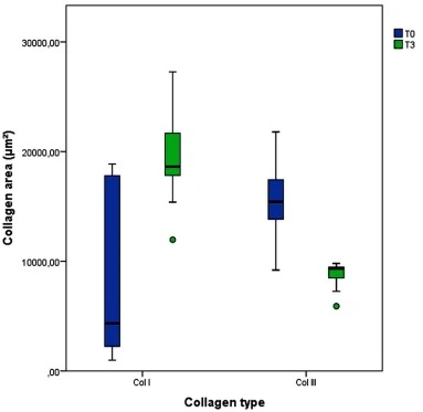
Fig. 3. Boxplot of type I and type III collagen levels at baseline (T0) and post-treatment (T3) in the study group (n = 9). The paired Wilcoxon test showed a significant increase in type I collagen (p = 0.038) and a reduction in type III collagen (p = 0.017) after treatment.
Morphologically, collagen bundles at T3 appeared thicker, longer, and more continuous, arranged in compact fascicular patterns with reduced interstitial spaces. Additionally, more intense acidophilic staining was observed in the periadnexal regions and deep reticular dermis. It is noteworthy that, although fiber organization was substantially modified, total collagen density did not show a statistically significant difference between periods (p = 0.744), suggesting that the main observed effect was related to collagen fiber reorganization rather than an overall quantitative increase in the matrix.
Discussion
Following the intervention, a significant improvement was observed in all patient-reported satisfaction parameters, including perceived laxity, skin appearance, firmness, and comfort with genital appearance. The statistical significance demonstrated by the Wilcoxon test reinforces the consistency of these findings.
Pinch Test results corroborated the subjective findings, showing a progressive reduction in measurements after baseline, as demonstrated by the Friedman test. Pairwise comparisons indicated significant reductions between baseline and subsequent time points, suggesting improvement in tissue tone and elasticity, with possible maintenance of the therapeutic effect throughout follow-up.
From a physiological perspective, the integrity of vulvar skin depends on the organization of the dermal extracellular matrix (ECM), composed mainly of type I and type III collagen and elastic fibers. With aging and estrogen decline, fibroblast activity and protein synthesis decrease, leading to ECM disorganization and loss of tissue tone. In this context, energy-based therapies have been investigated as minimally invasive alternatives capable of stimulating tissue regeneration through controlled heating of dermal and submucosal layers, promoting neocollagenesis and collagen reorganization [10].
Similar results have been reported with technologies such as fractional laser and radiofrequency, which are associated with improvement of vaginal mucosa, increased epithelial thickness, and reduction of symptoms such as dryness, dyspareunia, and mild urinary complaints [12], [13]. The findings of the present study are consistent with this body of literature.
Comparable clinical outcomes were described by Kim et al. [14], who evaluated radiofrequency-based thermal therapy in two sessions involving the entire vaginal wall for the treatment of laxity. The authors reported significant improvements in perceived laxity, sexual function, and reduction of sexual distress, with maintenance of benefits over follow-up and no relevant adverse events. These findings support the safety and efficacy of radiofrequency-based technologies, in agreement with the results observed in the present study.
Histological findings demonstrated a qualitative transition in vulvar dermal architecture following monopolar radiofrequency intervention. The significant increase in type I collagen (p = 0.038), associated with a reduction in type III collagen (p = 0.017), indicates that clinical benefits are not related to volumetric expansion of the matrix—given the stable behavior of total collagen (p = 0.74)—but rather to stromal maturation and reorganization.
The predominance of type III collagen at baseline (T0) is characteristic of tissues with lower tensile strength and greater compliance, commonly observed in aging and genital laxity [8]. Radiofrequency-induced heating is described as promoting collagen contraction and activating cellular processes involved in tissue remodeling, including heat shock protein (HSP) signaling and the progressive replacement of thinner fibers (type III) with thicker, more organized type I collagen bundles [15], [16].
The increase in the Col I/III ratio (from 0.33 to 1.77) at 90 days (T3) represents the most relevant statistical finding for understanding treatment efficacy and may be interpreted as an indirect marker of structural tissue reorganization. Similar results were reported by Vicariotto et al. [16], who observed an association between clinical improvement and collagen density following energy-based therapies. The absence of significant changes in total collagen suggests that radiofrequency primarily acts as a remodeling agent, optimizing the proportion between collagen isoforms to restore vulvar structural support.
This pattern of structural reorganization may explain the high levels of patient satisfaction, even in the absence of a marked increase in dermal thickness [5]. The fascicular organization observed at T3 provides biological support consistent with improved firmness and vulvar trophism, suggesting a positive structural impact on the ECM.
Despite these promising results, this study has limitations. The small sample size may have limited statistical power to detect quantitative variations in total collagen (p = 0.36), particularly considering the interindividual variability observed at baseline.
Nevertheless, significant changes were observed in matrix quality, particularly in the increase of type I collagen and the Col I/III ratio, suggesting that structural remodeling may represent an earlier biological effect of radiofrequency than volumetric matrix expansion. Future studies with larger samples and longer follow-up are needed to confirm the durability of these findings and to evaluate potential late changes in total collagen.
Conclusion
Non-ablative monopolar radiofrequency with automatic cooling promoted clinical improvement in vulvar laxity, associated with structural changes in the dermal extracellular matrix, characterized by increased type I collagen and an elevated collagen I/III ratio. Improvements in clinical and subjective outcomes reported by participants were also observed. These findings suggest that this approach represents a minimally invasive and potentially effective alternative for the management of vulvar laxity. Further studies with larger sample sizes and longer follow-up are required to confirm the durability of the observed effects.
Credit authorship contribution statement
Lourena Guedes de Melo Romão: Writing – review & editing, Writing – original draft, Validation, Methodology, Investigation, Data curation, Conceptualization. Ana Cristina de Moura Batalha: Writing – review & editing, Writing – original draft, Investigation, Data curation. Natália Gontijo Ribeiro: Writing – review & editing, Visualization, Formal analysis, Data curation. Willany Lauyne Ananias Mesquita: Writing – review & editing, Writing – original draft, Validation, Investigation, Formal analysis. Adriano Francisco Alves: Writing – review & editing, Writing – original draft, Validation, Methodology, Investigation, Formal analysis. Cynthia Germoglio Farias de Melo: Writing – review & editing, Writing – original draft, Validation, Supervision, Software, Methodology, Formal analysis, Data curation, Conceptualization.
Funding
No funding was received for this study.
Declaration of generative AI and AI-assisted technologies in the manuscript preparation process
During the preparation of this work, the authors used AI-assisted tools for language editing. After using these tools, the authors reviewed and edited the content as needed and take full responsibility for the content of the published article.
Declaration of competing interest
The authors declare that they have no known competing financial interests or personal relationships that could have appeared to influence the work reported in this paper.
References
- [1]S. Palacios, et al.Structure of epithelial and stromal compartments of vulvar and vaginal tissueJ Sex Med, 15 (7) (2018), pp. 1011-1018
- [2]A.M. Farrell, et al.Alterations in fibrillin as well as collagens I and III and elastin occur in vulval lichen sclerosusJ Eur Acad Dermatol Venereol, 15 (3) (2001), pp. 213-217
- [3]A. Marzec, et al.Collagen and elastin differences in vulvar tissue of women with vulvar dermatoses and healthy womenGinekol Pol, 94 (4) (2023), pp. 289-295
- [4]M.P. GoodmanFemale genital cosmetic and plastic surgery: a reviewJ Sex Med, 8 (6) (2011), pp. 1813-1825
- [5]G. Leibaschoff, et al.Radiofrequency for the treatment of vulvovaginal laxity: a reviewJ Cosmet Dermatol, 15 (3) (2016), pp. 214-220
- [6]Andrade AC. Radiofrequency não ablativa no rejuvenescimento vulvovaginal. Undergraduate thesis. Federal University of Paraíba; 2025.
- [7]N.S. Sadick, R.S. MulhollandA prospective clinical study evaluating the efficacy of radiofrequency treatment for skin tighteningDermatol Surg, 30 (4) (2004), pp. 503-507
- [8]C. Karcher, N.S. SadickA practical guide to rejuvenate the female genitaliaInt J Womens Dermatol, 2 (2) (2016), pp. 39-46
- [9]M. Krychman, et al.Effect of single-treatment, surface-cooled radiofrequency therapy on vaginal laxity and female sexual function: The VIVEVE I randomized controlled trialJ Sex Med, 14 (2) (2017), pp. 215-225
- [10]S. Salvatore, et al.Histological study on the effects of microablative fractional CO2 laser on atrophic vaginal tissue: an ex vivo studyMenopause, 22 (8) (2015), pp. 845-849
- [11]Carvalho F, et al. Tratado de ginecologia regenerativa: estética e funcional. Rio de Janeiro: Fosthera; 2025.
- [12]M. Gambacciani, M. LevanciniVaginal erbium laser: Second-generation thermotherapy for GSMClimacteric, 18 (5) (2015), pp. 757-763
- [13]D.J. Portman, M.L.S. GassGenitourinary syndrome of menopause: new terminology for vulvovaginal atrophy Menopause, 21 (10) (2014), pp. 1063-1068
- [14]J.H. Kim, K. Kim, S. Ahn, Y. Lee, J.S. Kim, D.H. Suh, et al.Pilot study of radiofrequency thermal therapy performed twice on the entire vaginal wall for vaginal laxityEur J Obstet Gynecol Reprod Biol, 254 (2020), pp. 159-163, 10.1016/j.ejogrb.2020.09.022
- [15]E. Dayan, A.J. Burns, R.J. Rohrich, S. TheodorouThe use of radiofrequency in aesthetic surgeryPlast Reconstr Surg Glob Open, 8 (8) (2020), p. e2861
- [16]F. Vicariotto, M. RaichiNon-ablative 4 MHz dual radiofrequency device for vulvovaginal rejuvenation: an objective assessmentJ Cosmet Laser Ther, 19 (3) (2017), pp. 182-188